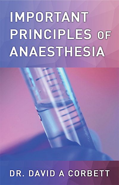
Important Principles of Anesthesia
$15.00
Students of medicine are required to learn a vast amount of information in a very short period.
Teachers seem to spend little time showing the relationship between theory and its application so
that a student has no perspective as to what is important and what is not.
Description
Part -1
The purpose of ventilation is to transfer oxygen from the external atmosphere to the alveoli and to remove carbon dioxide from the alveoli. It is a prerequisite for this that there should be a clear pathway between the oxygen supply and the alveoli.
Spontaneous ventilation
When patients breathe by themselves under a general anaesthetic , they will be spontaneously ventilating – with the proviso that they have a clear airway.
Whenever a patient is breathing spontaneously through an anaesthetic circuit, one must
observe two areas in particular:
- the movement of the reservoir bag and
- the tissues at the root of the neck.
The reservoir bag can be moving without the patient getting adequate ventilation. If the anaesthetic is too deep, they will not be moving their respiratory muscles adequately. But the commonest cause of inadequate ventilation is partial airway obstruction. In some cases (for example, a large patient), the reservoir bag may seem to have adequate excursion but the excursion is inadequate for that patient. If you do not observe the root of the neck and note indrawing of tissues during inspiration, you will probably be unaware that there is any obstruction at all.
“Noisy breathing is obstructed breathing”.
Controlled ventilation
Controlled ventilation must deliver adequate volumes of oxygen to the alveoli and allow adequate time for the removal of carbon dioxide. When using Positive End-Expiratory Pressure (PEEP) or Continuous Positive Airways Pressure (CPAP), you are performing a balancing act. Although you are enhancing the uptake of oxygen, you are also hindering the removal of carbon dioxide.
It is crucial that you monitor the following three factors:
- Upper airway pressure
The aim of upper airway pressure is to deliver adequate volumes of gas to the alveoli. The pressure must be high enough to do this. A pressure of 40 Torr that is not expanding the lungs is an inadequate pressure. One concern about high airway pressures is that the pressure might damage the alveoli (leading to barotrauma). In reality, it is only when excess volume is introduced
into the alveoli that damage can occur. Therefore, no matter how high the pressure reads on the ventilator, if this pressure is not introducing volume into the lungs then barotrauma to alveoli cannot occur (even though it might do damage to the trachea or produce surgical emphysema).
In asthmatics and patients with Chronic Obstructive Pulmonary Disease (COPD), higher airway pressures will be necessary to overcome airway resistance. These patients will also need a longer expiratory time to allow for the adequate removal of carbon dioxide.
Patients with flail chests must have their ventilators set to overcome any paradoxical movement of the flail segment. If the flail segment moves in during inspiration, then the intra-thoracic pressure must be sub-atmospheric at that point. Hence the airway pressure must be set high enough to introduce enough volume into the lungs to splint the flail segment.
- Inspiratory volume
This must be enough to deliver adequate volumes of oxygen to the alveoli and to remove adequate volumes of carbon dioxide from them. The volume is usually about 70–100 ml/kg/minute, with a tidal volume of about 7–10 ml/kg.
- Respiratory rate
In normal fit patients, we aim to have an expiratory time about twice that of the inspiratory time, and a ventilatory rate of about 6–15 times per minute (with 10 breaths/minute the easiest figure to calculate with when setting a ventilator).
Part 2
Because the hydration numbers of the cations decrease as the atomic number increases, Cesium, with atomic number of 55, is less hydrated than any of the above- mentioned cations. Cesium, which is also a univalent cation, can therefore displace Sodium and Potassium from receptor sites and bind more strongly to them. One might be tempted to ask what possible relevance Cesium might have to our discussion. It so happens that when nuclear power stations blow up (as occurred in Chernobyl
in 1986), they liberate large quantities of Cesium-137, which is radioactive. This is a substance which binds strongly to human tissues, particularly muscle, at the expense of the normal ions. Its half-life is about 30 years and it greatly increases the likelihood of cancer.
Another radioactive element of importance is Strontium-90. This, like Cesium-137, is a by-product of
nuclear fission. Strontium-90 is a divalent cation and will tend to displace Calcium and become deposited in bone and bone marrow. It also has a half-life of about 30 years.
Potassium ion
Let us now consider the intracellular environment. If there are negative receptor sites within the cell (and there are many of them on amino acids), which ion will preferentially bind to them? From the above discussion, Potassium will bind in preference to Sodium, but Hydrogen, if enough is available in free form, will displace Potassium. As Potassium can bind more strongly to intracellular
receptors than Sodium, this explains why Potassium, and not Sodium, is the major intracellular ion.
In body fluids, there are two forms of any given ion: a free and a bound form. The bound form is that bound to negative receptor sites (such as those on proteins) and the free form is that which is unbound. Within the cell, the overall concentration of potassium is about 150 mmol/L. If 145 mmol/L is bound to negative receptors, this leaves only 5 mmol/L in the free form. The
extracellular level of potassium is also about 5mmol/L.
Thus we have no real concentration gradient across the cell membrane. This is in line with the observation that Potassium (and Sodium, for that matter) moves freely across cell membranes depending on the direction of its concentration gradient
Part 3
A further point of interest is that the drainage of such a fountain-like system would require two venules (one for each hemisphere of capillaries) for each supplying arteriole. The reason for the double drainage becomes obvious when we consider what would happen if there were only one drainage vessel. In such a case, if the arteriole compressed and obstructed the only drainage venule, tissue oedema would result. Conversely, if the venule dilated and obstructed the arteriole, local gangrene would be the result.
Indeed the vascular system of the body does, as a
general rule, have two veins accompanying each artery
(venae comitantes). This is also true of the aorta where the
inferior vena cava is one of its venae comitantes while the
degenerative hemi-azygous veins on the left side completes
the other.
There is, however, one apparent exception: there are two umbilical arteries and only one umbilical vein in the foetus. But remember that the supply vessel to the foetus is the umbilical vein. So although there are two arteries histologically, these are, in fact, drainage vessels for the foetus. Thus, the system remains consistent: one supply vessel to a tissue accompanied by two drainage vessels.
Initially, the micro-circulation is closed due to the tone of the pre-capillary sphincter. As the tissue continues to metabolise, it exudes potassium and other waste products (which eventually renders the pre-capillary sphincter flaccid). The pressure of the circulation can now overcome the tone of the pre-capillary sphincter and blood is able to enter the capillary bed. This circulation removes the
potassium and other waste products of metabolism and replenishes the necessary oxygen and nutritional substances. The pre-capillary sphincter thus regains its original tone
and again closes off the circulation.
In this way, a tissue derives a circulation depending on its metabolic activity. A tissue with a higher metabolic activity will derive a greater share of the circulation than a tissue of lesser metabolic activity. Hence malignant tissue will supply itself with an excess amount of nutrient at the expense of normal tissue. The tumour will grow and the patient will become cachectic. This might also explain the common observation that a malignant tumour will often expand rapidly after a general anaesthetic. During a general anaesthetic, sympathetic nerves to pre-capillary sphincters and the musculature of the sphincters themselves also become anaesthetized and therefore allow an increased blood flow to all capillary beds.
Neural control
The pre-capillary sphincters are supplied by sympathetic fibres from the autonomic nervous system. These fibres maintain a certain basal tone in the sphincters. If a particular tissue begins to derive an increased proportion of blood volume (for example, to striated muscle as a result of increased exercise), pressure and volume receptors in the great vessels notify the brain that a circulatory deficiency is occurring. The brain increases its sympathetic output to produce a generalised increase in vasomotor tone. Thus the pre-capillary sphincters of less active tissues (e.g. the gut) are kept closed longer to divert the irrigation to the tissues in need.
This same mechanism acts when a deficit in intravascular volume occurs (for example, as a result of haemorrhage, burns or dehydration). A generalised increase in vasomotor tone results. Skin, gut and kidney therefore have their vascular supplies reduced to allow adequate perfusion of the brain and heart. Thus we get pallor, nausea and decreased urine output. (Decreased urinary output also conserves intravascular volume.)
With increasing sympathetic output from the brain, the tone of the pre-capillary sphincters is increased. Thus the metabolic waste products from the cells have to reach ever higher levels to overcome this tone before the cells can derive nutrition for themselves. But the higher levels of
Available at :
*Price may vary by retailer
Get a FREE ebook by joining our mailing list today!
Lorem ipsum dolor sit amet, consectetur adipiscing elit.


Reviews
There are no reviews yet.